Home » Without Label » Upper Back Anatomy : Not-So-Gross Anatomy: Lats & Upper Back — b3 Wellness : The anatomy of the back refers to the muscles of the back, as well as the bones of the scapulae, ribcage, and spine.
Upper Back Anatomy : Not-So-Gross Anatomy: Lats & Upper Back — b3 Wellness : The anatomy of the back refers to the muscles of the back, as well as the bones of the scapulae, ribcage, and spine.
Upper Back Anatomy : Not-So-Gross Anatomy: Lats & Upper Back — b3 Wellness : The anatomy of the back refers to the muscles of the back, as well as the bones of the scapulae, ribcage, and spine.. All these muscles are therefore associated with movements of the upper limb. The hurt can stem from sore muscles, ligaments, and tendons, or from herniated disks, fractures, and other problems in your upper, middle, and lower back. In the upper back region, the trapezius, rhomboid major, and levator scapulae muscles anchor the scapula and clavicle to the spines of several vertebrae and the occipital bone of the skull. See human back anatomy stock video clips. They originate from the vertebrae and insert into the scapulae.
The iliocostalis muscles are furthest from the spine. Latissimus dorsi (lats), the largest muscle in the upper part of your body. The bones of the chest and upper back combine to form the strong, protective rib cage around the vital thoracic organs such as the heart and lungs. In the upper back region, the trapezius, rhomboid major, and levator scapulae muscles anchor the scapula and clavicle to the spines of several vertebrae and the occipital bone of the skull. The upper back has the most structural support, with the ribs attached firmly to each level of the thoracic spine and very limited movement.
1000+ images about MUSCLE ANATOMY on Pinterest | Back pain ... from s-media-cache-ak0.pinimg.com This muscle is located on the upper portion of the back anatomy, underneath the trapezius. They originate from the vertebrae and insert into the scapulae. 3d human upper leg anatomy or anatomical and muscle set or collection. These muscles facilitate movement by attaching to one or. The trapezius and latissimus dorsi muscles connect the upper limb to the vertebral column. The deltoid, teres major, teres minor, infraspinatus, supraspinatus (not shown) and subscapularis muscles (not shown) all extend from the scapula to the humerus and act on the shoulder joint. The superficial back muscles are situated underneath the skin and superficial fascia. The trapezius and latissimus dorsi muscles connect the upper limb to the vertebral column.
The cervical spine supports the weight and movement of your head and protects the nerves exiting your brain.
Human body anatomy female female anatomy muscle shoulder blade pain anatomy back muscles bones man female anatomy body muscles in a body female anatomy muscole shoulder concept muscular sysyem. Back muscles anatomy here include the trapezius, latissimus dorsi, rhomboid and levator scapulae. The iliocostalis muscles are furthest from the spine. It consists of seven vertebrae. Covering an expanse from the neck to the tailbone, the back muscles are responsible for a broad range of functions, from extending the spine to shrugging the shoulders. Because the upper back was designed to be strong, inflexible and incredibly stable, it's difficult to injure this area or for this area of the back to degenerate over time. There is a set of muscles in the upper back (called the thoracic area) called the spinalis thoracis. Anatomy of the upper back muscles. In the upper back region, the trapezius, rhomboid major, and levator scapulae muscles anchor the scapula and clavicle to the spines of several vertebrae and the occipital bone of the skull. They originate from the vertebrae and insert into the scapulae. The teres majo r muscles work with the rotator cuff muscles to stabilize. The neck consists of seven cervical vertebrae, the building blocks of the spine. The intricate anatomy of the back provides support for the head and trunk of the body, strength in the trunk of the body, as well as a great deal of flexibility and movement.
In the upper back region, the trapezius, rhomboid major, and levator scapulae muscles anchor the scapula and clavicle to the spines of several vertebrae and the occipital bone of the skull. The upper back has the most structural support, with the ribs attached firmly to each level of the thoracic spine and very limited movement. The cause may be poor posture (such as forward head posture) or any type of irritation of the large back and shoulder muscles, including muscle strain or spasms. The anatomy of the back refers to the muscles of the back, as well as the bones of the scapulae, ribcage, and spine. They originate from the vertebrae and insert into the scapulae.
Neck And Upper Back Muscles Anatomy - ANATOMY STRUCTURE from lh6.googleusercontent.com Latissimus dorsi (lats), the largest muscle in the upper part of your body. This is my video about the muscles of the back. Powerful muscles that move the head and arms attach to these bones as well. So since we identified what we think are some of the best upper back exercises you can perform to fully develop your upper back, let's put these into a workout! 3d human upper leg anatomy or anatomical and muscle set or collection. Covering an expanse from the neck to the tailbone, the back muscles are responsible for a broad range of functions, from extending the spine to shrugging the shoulders. Both the deltoid and the trapezius are firmly attached to the spine of the scapula. This muscle is located on the upper portion of the back anatomy, underneath the trapezius.
Upper back pain is usually due to strained muscles, joint dysfunction, or a herniated disk, although these things are very rare.
In the upper back region, the trapezius, rhomboid major, and levator scapulae muscles anchor the scapula and clavicle to the spines of several vertebrae and the occipital bone of the skull. Human body anatomy female female anatomy muscle shoulder blade pain anatomy back muscles bones man female anatomy body muscles in a body female anatomy muscole shoulder concept muscular sysyem. The seventh cervical vertebra, referred to as c7, meets the first of 12 thoracic vertebrae t1 at the base of the neck, a. The thoracic spine —also referred to as the upper back or middle back—is designed for stability to anchor the rib cage and protect vital internal organs within the chest. The deltoid, teres major, teres minor, infraspinatus, supraspinatus (not shown) and subscapularis muscles (not shown) all extend from the scapula to the humerus and act on the shoulder joint. There is a set of muscles in the upper back (called the thoracic area) called the spinalis thoracis. The upper back has the most structural support, with the ribs attached firmly to each level of the thoracic spine and very limited movement. Both the deltoid and the trapezius are firmly attached to the spine of the scapula. The rhomboid muscle is activated as you bring and squeeze your scapula or shoulder blades back and together. Vertebrae there are 12 vertebrae in the thoracic spine. It is very stiff, and the thoracic spine has a limited range of motion. The upper back is a complex area containing a number of muscles that perform various actions on the scapulae (shoulder blades) and humerus. The iliocostalis muscles are furthest from the spine.
The upper back has the most structural support, with the ribs attached firmly to each level of the thoracic spine and very limited movement. Vertebrae there are 12 vertebrae in the thoracic spine. The iliocostalis muscles are furthest from the spine. See human back anatomy stock video clips. See upper back stock video clips.
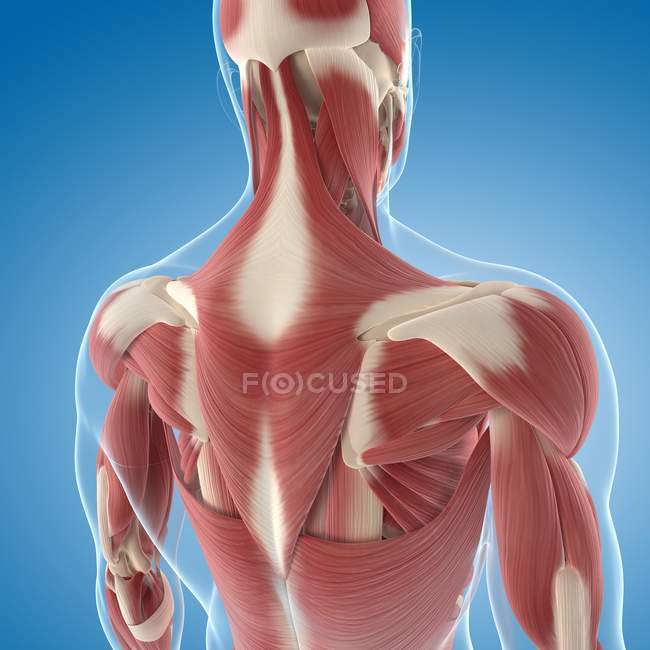
Upper Back Anatomy : Posterior Anatomy Upper Back Diagram ... from st.focusedcollection.com Powerful muscles that move the head and arms attach to these bones as well. Upper back pain is most commonly caused by muscle irritation or tension, also called myofascial pain. Latissimus dorsi (lats), the largest muscle in the upper part of your body. The cause may be poor posture (such as forward head posture) or any type of irritation of the large back and shoulder muscles, including muscle strain or spasms. It is very stiff, and the thoracic spine has a limited range of motion. The intricate anatomy of the back provides support for the head and trunk of the body, strength in the trunk of the body, as well as a great deal of flexibility and movement. In the upper back region, the trapezius, rhomboid major, and levator scapulae muscles anchor the scapula and clavicle to the spines of several vertebrae and the occipital bone of the skull. Both the deltoid and the trapezius are firmly attached to the spine of the scapula.
The thoracic spine —also referred to as the upper back or middle back—is designed for stability to anchor the rib cage and protect vital internal organs within the chest.
The bones of the chest and upper back combine to form the strong, protective rib cage around the vital thoracic organs such as the heart and lungs. In the upper back region, the trapezius, rhomboid major, and levator scapulae muscles anchor the scapula and clavicle to the spines of several vertebrae and the occipital bone of the skull. Upper back pain is usually due to strained muscles, joint dysfunction, or a herniated disk, although these things are very rare. This area of the body includes the top of the thoracic spine, which. A sample upper back workout. More commonly known as the shoulder blade, the scapula is a flat triangular bone located in the upper back. Sometimes you feel the effects right away. In the upper back region, the trapezius, rhomboid major, and levator scapulae muscles anchor the scapula and clavicle to the spines of several vertebrae and the occipital bone of the skull. The upper back has the most structural support, with the ribs attached firmly to each level of the thoracic spine and very limited movement. Because the upper back was designed to be strong, inflexible and incredibly stable, it's difficult to injure this area or for this area of the back to degenerate over time. Back muscles anatomy here include the trapezius, latissimus dorsi, rhomboid and levator scapulae. The trapezius and latissimus dorsi muscles connect the upper limb to the vertebral column. Anatomy of the upper back.